In order to have your best and healthiest smile, your dentists need to be able to detect problems before they escalate. Our skillful practitioners will be able to see signs of decay, oral cancer, and gum disease within a few minutes. Part of this ability comes from extensive training, and the other part comes from innovative technology!
What are Digital X-rays?
Digital x-rays show us images of every area of your teeth and mouth. Of course, our dentists can’t see between your teeth or inside the teeth with a visual inspection, but digital x-rays make that possible!
We use our digital x-ray to:
- Take images of your teeth from different angles
- Check for hiding decay, bacteria, disease, orthodontic issues, tumors, and oral cancer signs
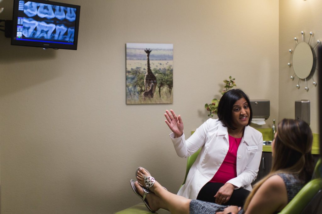
- Help the patient see the structures of their mouth and understand their oral health
- Provide an accurate and honest diagnosis
- Tailor a detailed treatment plan that will give you the best smile results
Digital x-rays have important functions, and also offer many benefits to the patients. Some of the advantages of digital x-rays over traditional x-rays include:
- Significantly reduced radiation exposure
- Instant image viewing–no waiting for images to develop
- Easy to transfer and save images for insurance acceptance
What is an Intraoral Camera?
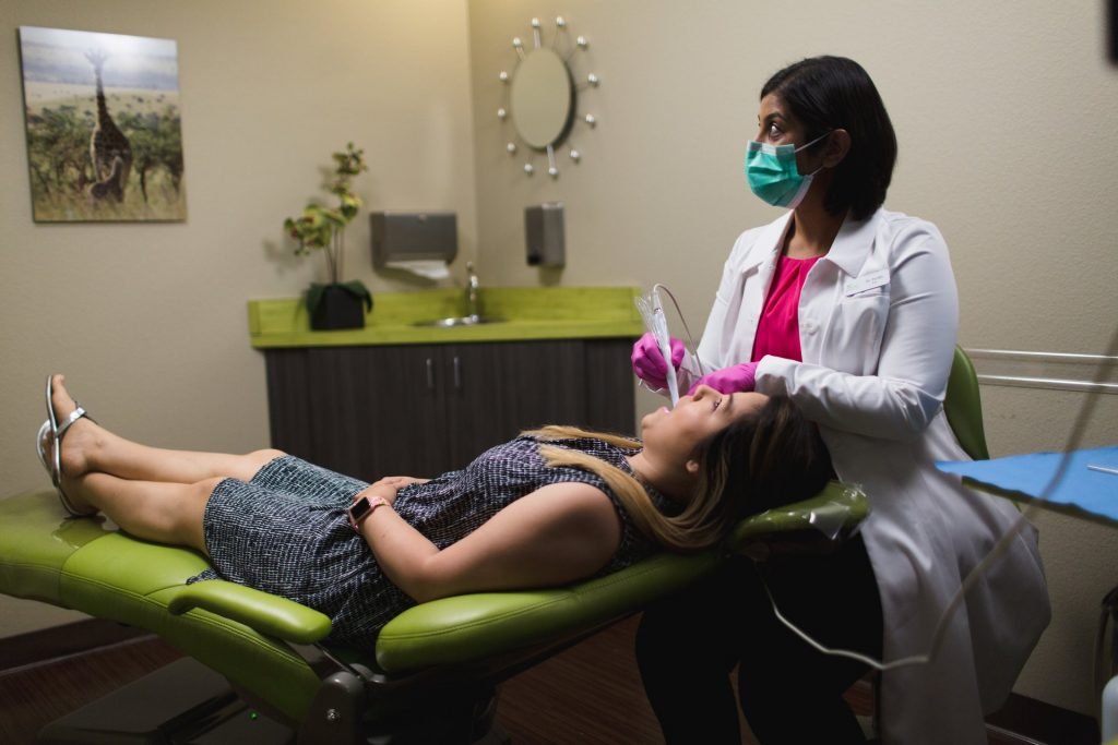
Intraoral cameras also show aspects of the teeth that are not visible to the naked eye. Instead of depicting the insides of the teeth like digital x-rays do, intraoral cameras give us detailed images of the exterior teeth.
An intraoral camera is a pen-sized device that is comfortable for the patient. Intraoral cameras allow your dentist to see and take photos of every angle of your mouth. An intraoral camera has extraordinary abilities, including:
- Transmitting real-time images of the teeth
- Zooming in on and highlighting different areas
You deserve precise diagnoses and treatments, and we make sure of that with advanced technology. To schedule an exam in the greater Fort Worth area, contact Mint Leaf Dental today. We look forward to working with you and treating you to excellent dentistry!
Isodry®
Have you ever experienced fatigue at a dentist’s office while trying to keep your mouth open during treatment? If so, we have some great news for you!
Dr. Parikh uses a breakthrough dental device called Isodry® that will make your next cleaning with us quite comfortable! Isodry’s flexible, single-use rubber fitting helps keep your mouth comfortably open, while an advanced suction tube system makes cotton rolls and suction wands a thing of the past!
Intraoral Scanner
An intraoral scanner is a handheld dental device that creates fast, comfortable, and highly accurate 3D digital impressions of your teeth, replacing messy traditional putty molds for crowns, clear aligners (like Invisalign), bridges, implants, and even monitoring oral health, allowing for better-fitting restorations, instant visualization of issues, and improved communication with your dentist. It works by taking thousands of photos that stitch together into a detailed model, making procedures more precise, faster, and less uncomfortable.
What it is
A small, wand-like device with a light and camera that scans your mouth. Captures detailed digital images, not physical molds.
How it Works
The scanner moves over your teeth and gums, capturing numerous images. Software stitches these images into a precise 3D model displayed on a screen. This digital file is sent instantly to a lab.
Benefits for patients
- Comfort: No more goopy impression material or trays in your mouth.
- Accuracy: High-resolution images lead to better-fitting crowns, bridges, and implants.
- Speed: Faster process and quicker turnaround times for lab work.
- Visualization: You can see your own teeth and potential problems on screen, helping you understand treatment plans.
- Versatility: Used for orthodontics (Invisalign), implants, crowns, bridges, and monitoring changes over time.
What to expect during a scan
A painless, quick process (often 5-10 minutes). You’ll feel the scanner moving, but it’s non-invasive and doesn’t involve radiation. You may be asked to bite down to capture your bite perfectly.
Cone Beam CT (CBCT)
Cone Beam CT (CBCT) in dentistry is a 3D X-ray that gives detailed views of teeth, bone, nerves, and soft tissues, used when standard X-rays aren’t enough for complex treatments like implants or wisdom teeth removal. It’s quick, painless, non-invasive, uses less radiation than hospital CTs, and involves standing or sitting still in the machine as it rotates around your head. This technology provides your dentist with precise information for better diagnosis and treatment planning.
What it is & Why it’s Used
- 3D Imaging: Creates detailed 3D models of your mouth, jaws, sinuses, and nerves.
- Better Than 2D: Reveals structures obscured on flat X-rays, improving diagnostic accuracy for complex issues.
- Common Uses: Planning dental implants, complex extractions (impacted wisdom teeth), root canals, orthodontics, and diagnosing pain/disease.
What to Expect During the Scan
- Positioning: You’ll stand or sit, bite on a piece of plastic, and may have your head held still with straps.
- During the Scan: The machine rotates around your head; it’s quick (seconds) and quiet, unlike an MRI.
- Stay Still: Keeping still is crucial for sharp images.
- No Sedation: Usually not needed as it’s painless.
Before Your Scan
- Preparation: Little to none, but wear comfortable clothes and remove jewelry.
- Pregnancy: Inform your dentist if you might be pregnant.
- Claustrophobia: Tell your dentist if you’re prone to claustrophobia so they can help.
After the Scan
- Immediate Return: You can resume normal activities right away.
- Review: Your dentist will show you the 3D images to discuss your treatment plan.
Safety & Radiation
- Lower Dose: Uses significantly less radiation than traditional medical CT scans.
- Necessity: Dental X-rays, including CBCT, should only be performed when necessary, so discuss benefits/risks with your provider.